Biological macromolecules are highly susceptable to radiation damage when exposed to the electron beam. Therefore, imaging macromolecules using the electron microscope entails some kind of averaging over molecule images that have been obtained with an extremely low dose (low-dose imaging). For such an average to be meaningful, the molecules must be in precisely the same conformation and binding state.
Traditionally, to obtain multiple images of an entire ensemble of molecules, one would use a crystal. In a crystal, all molecules are in the same conformation and binding state (as a result of high purification that is normally a prerequisite to crystallization, and of the highly homogeneous microenvironment of each molecule). They are also, convenient for averaging, in the same orientation and ordered position. X-ray crystallography makes use of the high order, since the ordered arrangement enables the collection of diffraction patterns. In electron microscopy, the strong interaction of electrons with matter implies that the crystal should be no thicker than a single layer (two-dimensional crystal}.
Advantages of using two-D crystals:
Disadvantages:
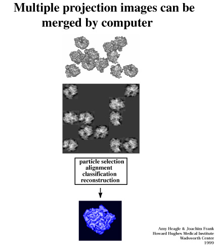
Single-particle reconstruction is the term used for the reconstruction of a macromolecule from images of a specimen in which the molecule exists in many realizations in the form of single, isolated particles, i.e., without contact with neighboring molecules. Thus, without the need for crystallization, there is in principle no restriction on the kinds of macromolecules that can be reconstructed.
Advantages of using single-particle reconstruction: