What is the contrast transfer function?
The image formation in bright field electron microscopy can be described by the action of the contrast transfer function (CTF) H(k). Accordingly, the relationship between the object o(r) and the image contrast i(r) can be written as i(r) = o(r)* h(r), where * stands for the convolution operation, and h(r) is the point spread function, which is the Fourier transform of H(k). Thus, following the convolution theorem, I(k) = O(k)H(k).
The shape of the CTF, H(k), depends on several parameters (for details, see Frank, 2006):
defocus [A] - which describes the deviation in the focus of
the objective lens from the "Gaussian focus."
spherical aberration coefficient [mm] - which describes the
(third order) spherical aberration of the wave front in the objective lens.
source size [1/A] - which describes the illumination divergence,
expressed as a size in the back focal plane (hence a quantity in
reciprocal space).
defocus spread - which describes the spread of defocus due to
the spread of electron energies or to the fluctuation of lens current.
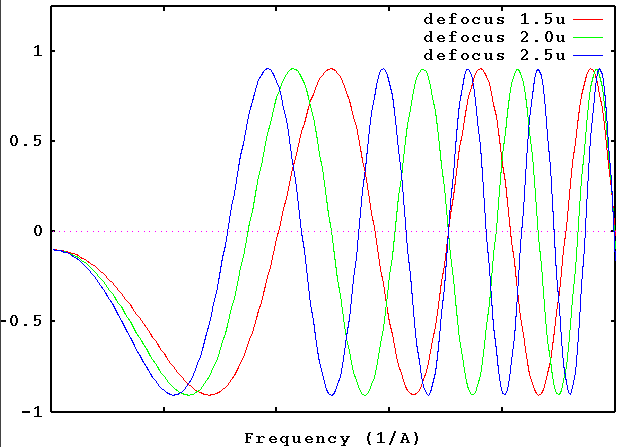
The only parameter being varied in the experiment is the defocus. Depending on the defocus setting, different features of the object appear enhanced or suppressed in the image. This is because the CTF oscillates between -1 (negative contrast transfer) and +1 (positive contrast transfer) as we go from low to high spatial frequencies. The exact locations of the zero crossings (where no contrast is transferred, and information is lost) depends on the defocus.
Contrast transfer functions - 3 defocus settings
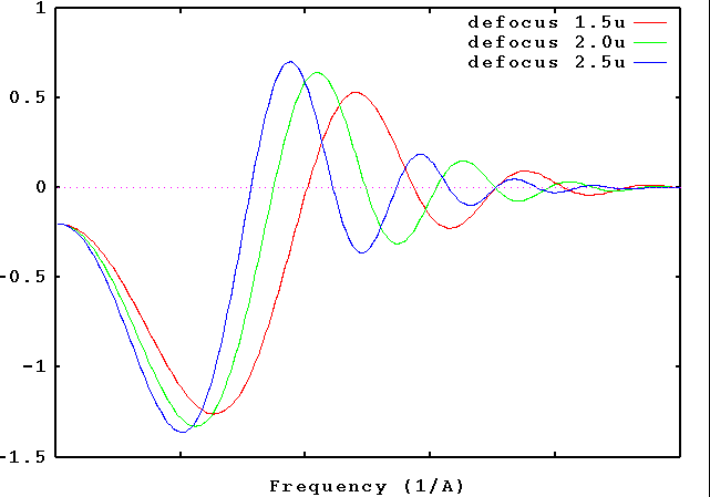
Contrast transfer functions - effect of envelope functions
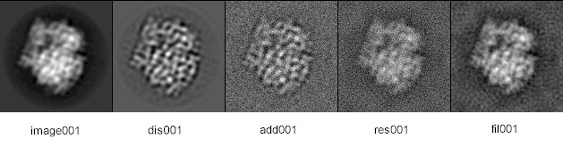
In CTF correction, we attempt to retrieve the undistorted object from the image. This attempt is compromised by the presence of noise; i.e., recovery of the object is never ideal. Since the CTF always has zero crossings, part of the information about the object is lost. This is why we make use of several images obtained at different defocus settings, hoping that the resulting CTFs Hn(k) jointly (after appropriate weighting) cover the whole Fourier space without gap.
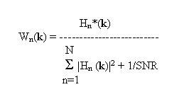
The Wiener filter is the least square solution to the problem of signal recovery in the presence of noise. Let's assume we have N images in (r) (with Fourier transforms In(k)) whose CTFs are Hn(k). In that case, the best estimation of the object transform O(k) is
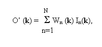 where
where